By Sunil Bochare MD, Melissa Nuncio FNP
University Health, San Antonio, Texas
sunil.bochare@uhtx.com
A previously healthy 5-year-old girl presents to clinic accompanied by her mother for evaluation of a persistent cough ongoing for three weeks. Over the past three days, she has developed subjective fevers, watery diarrhea, and facial swelling. Her mother reports that the cough is severe and comes in fits, causing the child to turn red in the face. The child has no significant past medical history and has not been seen by a primary care provider regularly. Her immunizations were reported to be up to date until two years of age. The mother also notes that multiple family members have had similar upper respiratory symptoms over the past few weeks. There is no history of vomiting, trauma, or foreign body aspiration.
At the time of presentation, the child is afebrile, alert, and interactive but appears fatigued. Vital signs are notable for mild tachycardia and normal blood pressure. Oxygen saturation is 95% on room air. Physical examination reveals a soft, non-erythematous swelling over the right cheek and lateral neck. Palpation elicits crepitus. Respiratory examination demonstrates diffusely decreased breath sounds bilaterally without wheezing or rales. There are no signs of respiratory distress such as nasal flaring, retractions, or cyanosis.
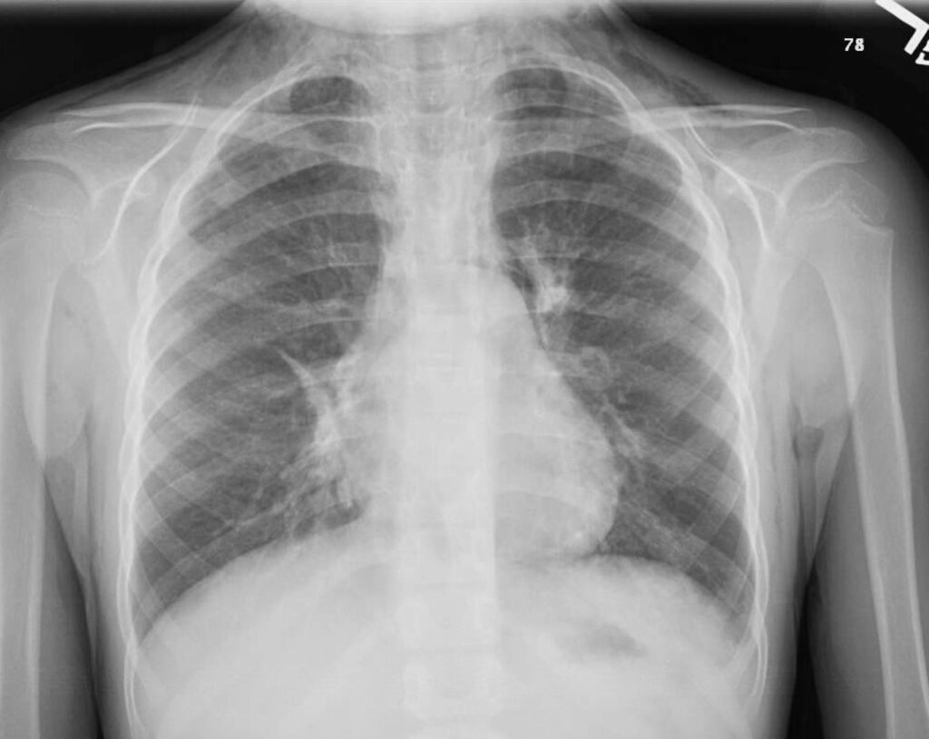
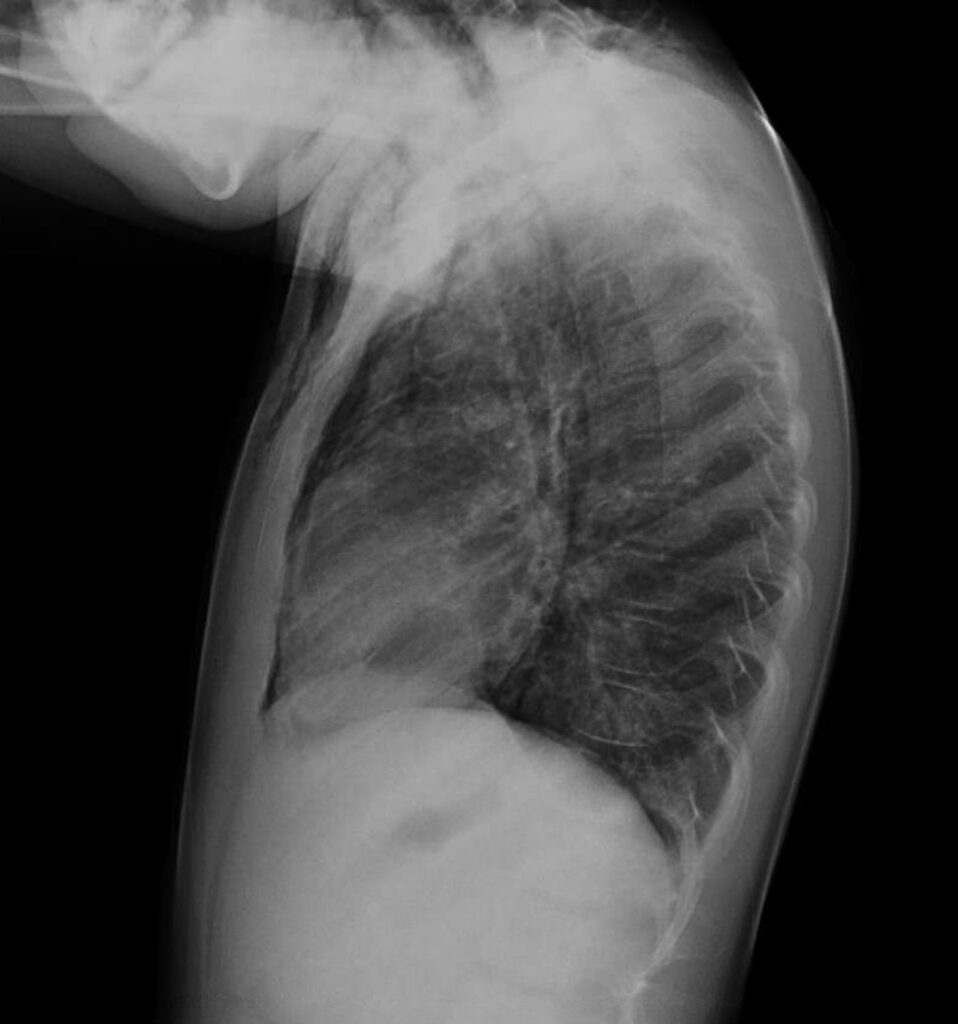
Given the concern for soft tissue emphysema, a chest X-ray is obtained. Imaging reveals abnormal lucency in the mediastinum with air tracking into the neck and bilateral chest wall. Bronchial wall thickening and streaky opacities in the right middle lobe are also noted. No pneumothorax or pleural effusion is seen. The child is referred urgently to the emergency department for further evaluation and advanced imaging.
Differential diagnosis at this point
- Subcutaneous emphysema secondary to trauma
- Retropharyngeal abscess
- Necrotizing pneumonia
- Esophageal perforation (Boerhaave syndrome)
Actual Diagnosis
- Spontaneous pneumomediastinum associated with respiratory syncytial virus (RSV) infection.
Discussion
Spontaneous pneumomediastinum (SPM) is defined as the presence of air within the mediastinum without a precipitating traumatic or iatrogenic event. It is rare in the pediatric population, with an estimated incidence of 1 in 30,000 emergency department visits. The most common age range for pediatric SPM is between 6 and 17 years, making cases in younger children less frequently encountered. While the condition is often benign, its dramatic clinical and radiographic presentation may raise concern for more serious pathology.
The pathophysiology of SPM centers on the Macklin effect, a mechanism first described in 1939. The sequence involves alveolar rupture due to sudden elevation in intra-alveolar pressure, air leakage into the pulmonary interstitium, and subsequent dissection along the bronchovascular sheaths into the mediastinum. This phenomenon can be triggered by events such as forceful coughing, vomiting, asthma exacerbations, or Valsalva maneuvers.
Respiratory syncytial virus (RSV) is a ubiquitous pathogen in pediatric populations and a common cause of bronchiolitis and lower respiratory tract infections. Though air leak syndromes are more commonly associated with mechanical ventilation or underlying pulmonary disease, they can occur in immunocompetent, otherwise healthy children, particularly in the setting of intense coughing spells, as seen in this case. Rarely, RSV-associated coughing has been implicated in pneumomediastinum and even spontaneous pneumothorax.
The clinical presentation of SPM varies with the extent of air dissection. Common symptoms include chest pain, dyspnea, dysphonia, neck swelling, facial puffiness, and subcutaneous crepitus. In children, particularly nonverbal or young patients, these symptoms may be vague or attributed to viral illness. A high index of suspicion is required when new or asymmetric swelling, hoarseness, or diminished breath sounds are detected during physical examination.
Chest radiography remains the first-line imaging modality and is diagnostic in most cases. Radiographic signs of pneumomediastinum include linear lucencies outlining mediastinal structures, air surrounding the heart and great vessels, and air tracking into the soft tissues of the neck. In uncertain or complicated cases, especially when esophageal or tracheal injury is a consideration, computed tomography (CT) provides enhanced anatomical detail. Contrast-enhanced CT esophagram can help rule out esophageal perforation, which is a life-threatening diagnosis that must not be missed.
In this case, CT of the chest and neck confirmed the presence of pneumomediastinum dissecting into the upper facial and cervical soft tissues. No evidence of contrast extravasation was seen, effectively ruling out esophageal rupture. A small right-sided perifissural pneumothorax and patchy consolidative opacities were also noted, raising concern for a superimposed infectious or inflammatory process. Respiratory viral panel returned positive for RSV, and no other pathogens were detected. Laboratory tests including procalcitonin were within normal range, supporting a viral rather than bacterial etiology.
The mainstay of treatment for uncomplicated SPM is conservative management. Supplemental oxygen can help facilitate nitrogen washout and accelerate the reabsorption of free air. Analgesia, antipyretics, and hydration support are generally sufficient. Avoidance of maneuvers that increase intrathoracic pressure, such as forceful coughing, is advised. Antibiotics are typically reserved for cases with clinical or radiologic evidence of secondary bacterial infection.
Hospitalization may be necessary for patients with hypoxia, respiratory distress, or comorbid conditions. The decision to monitor in an inpatient setting depends on the clinical stability of the patient and the presence of complications such as pneumothorax or suspected airway injury. Serial examinations and imaging may be warranted to ensure resolution and to detect potential progression.
In the presented case, the patient was admitted due to her oxygen requirement and abnormal imaging findings. She received 1.5 L of nasal cannula oxygen, IV fluids, and empirical antibiotics (azithromycin and ceftriaxone) in the emergency department. Antibiotics were discontinued after RSV was confirmed and no evidence of bacterial infection emerged. She remained hemodynamically stable, with no escalation in oxygen needs, and gradually returned to her clinical baseline.
Patient Course
The patient remained clinically stable during hospitalization. Oxygen was weaned over 48 hours, and her oral intake improved with supportive care. Repeat imaging was not required due to steady clinical improvement. She was discharged home after three days with no ongoing respiratory symptoms and was instructed to follow up with her primary care provider.
Summary
- Pneumomediastinum is an uncommon complication of viral respiratory infections, including RSV, and should be considered in children with persistent or forceful coughing and new-onset neck or facial swelling.
- The Macklin effect is the underlying mechanism for spontaneous air dissection into the mediastinum and subcutaneous tissues.
- Key clinical signs include subcutaneous crepitus, diminished breath sounds, and chest or neck swelling.
- Imaging—starting with chest X-ray—is critical; CT may be needed to exclude serious etiologies like esophageal rupture.
- Management is largely supportive. Hospitalization is indicated for hypoxia, respiratory distress, or uncertain diagnosis.
- Education of families on red flags (e.g., worsening respiratory symptoms, fever recurrence) is essential to ensure timely reassessment.
References and Suggested Readings
- Sahni S, Verma S, Grullon J, Esquire A, Patel P, Talwar A. Spontaneous pneumomediastinum: time for consensus. N Am J Med Sci. 2013;5(8):460–464.
- Johnson JN, Jones R, Wills BK. Spontaneous pneumomediastinum. West J Emerg Med. 2008 Nov;9(4):217-8. PMID: 19561749; PMCID: PMC2672281.
- Takada K, Matsumoto S, Hiramatsu T, et al. Spontaneous pneumomediastinum: an algorithm for diagnosis and management. Ther Adv Respir Dis. 2009;3(6):301–307.
Figures